

Expert neurosurgeon shares insights on recent developments in the fight against the aggressive brain cancer
Glioblastoma Awareness Day 2022 brings attention to the critical importance of finding a cure for glioblastoma, the most aggressive form of brain cancer. This day serves as a tribute to patients, families, caregivers, clinicians, and researchers who tirelessly work towards improving the lives of those affected by glioblastoma. On this occasion, Dr Rahul Gupta, Director of Neurosurgery at Fortis Hospital, Noida, provides valuable insights into the advancements made in the treatment of glioblastoma over the past five years.
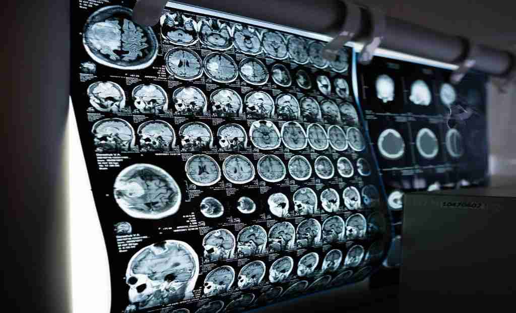
Pre-operative Assessment: Early Detection and Improved Localization
With the widespread availability of CT and MRI scans, the early detection of glioblastoma has become more feasible. This enables patients to undergo surgery at an early stage, leading to better outcomes. Furthermore, the development of new MRI sequences, such as spectroscopy, functional imaging, perfusion studies, and tractography, has significantly enhanced tumor localization and surgical planning. These advancements aid in determining the trajectory of surgery and the precise location of craniotomy, facilitating minimally invasive techniques. Additionally, pre-operative techniques such as DSA and embolization have proved beneficial for certain vascular skull base tumors.
Surgical Innovations for Safe and Complete Tumor Removal
- Operating Microscope: The utilization of high-end operating microscopes has become indispensable in brain tumor surgeries. These advanced devices magnify the tumor, enabling neurosurgeons to achieve safe and complete removal of brain tumors.
- Intraoperative Fluorescence: The introduction of intraoperative fluorescence techniques has revolutionized tumor localization during surgery. By using special dyes and filters through software integrated into the operating microscope, neurosurgeons can distinguish between normal brain tissue and tumor cells in real-time.
- Awake Craniotomy: In cases where tumors are located in eloquent areas of the brain, especially the dominant side, awake craniotomy has proven beneficial. This procedure involves sedating the patient while performing the craniotomy under local anesthesia. The patient is then asked to speak and move limbs on command during tumor removal. This approach minimizes post-operative deficits and improves overall outcomes.
- Neuro Monitoring: To prevent any damage to functionally important areas of the brain, neuro monitoring is employed. This technique involves checking the electrical activity of the brain to localize various functional regions. Electro-physiologists are present in the operation theatre to assist with this process.
-
Also Read: Max Healthcare and IIT Bombay Join Hands to Drive Healthcare Innovation