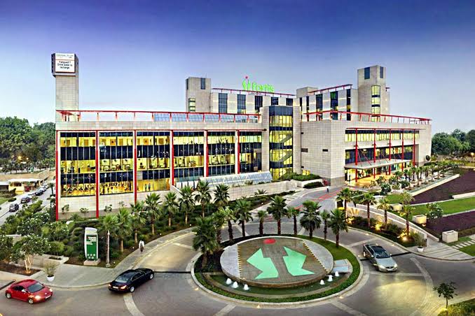
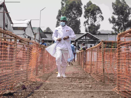
A team of doctors at Fortis Memorial Research Institute (FMRI), Gurgaon performed a lifesaving surgery on a 10-month-old boy who had a congenital condition known as ‘tracheal hemangioma.’ The condition is one where there is a huge constantly growing mass present in the airway making it impossible to breathe. The baby was managed by a team of clinicians led by Dr. Krishnan Chugh, Director& HOD, Paediatrics, FMRI and Dr. Anand Sinha, Senior Consultant, Paediatric Surgery, FMRI.
On presentation, the baby had severe difficulty in breathing. A thin flexible camera scope was passed into the baby’s airway to look for the cause. Dr. Krishnan Chugh said, “A large mass was growing within the baby’s trachea, which was blocking over 90% of the air passage. This mass or hemangioma is essentially an overgrowth of blood vessels within the trachea (airways). The baby was immediately started on Propranolol, a medicine which is often used to effectively treat the condition. However, before the drug could have any effect, the rapidly growing mass made breathing extremely difficult. He had to be put onto a mechanical ventilator. However, that proved to be ineffective in pushing the air beyond the growing mass. A surgical removal was planned, but the surgery itself was potentially life-threatening and there were several possible complications. However, the worsening condition of the baby left us with no choice.”
Dr. Anand Sinha, Consultant, Paediatric Surgery, FMRI said, “There were a lot of technical difficulties associated with the surgery. Accessing the mass in the tiny trachea of the baby was difficult, and there was a very high risk of bleeding and damage to the trachea that could have resulted in an air leak. A rigid tube with camera was introduced in the trachea and with the help of extremely thin and fine instruments, the mass was burnt off. The biggest challenge was how to ensure oxygen level of the body is not disrupted while operating. It was a very tricky task for the anesthetists. It was difficult to reach the mass in his tiny trachea and the entire mass couldn’t be dispensed in one go as the child’s critical status did not allow intervention for a long duration. However, in two attempts, 72 hours apart, the entire mass was removed. Thankfully everything went well.”
Thanking the hospital for the treatment, the mother of the baby said, “It was honestly the most frightening ordeal that we have had to go through. We weren’t sure if our baby would make it, but we placed all our faith in the doctors and are thankful to them for his recovery! He is breathing fine now, and we could not be more relieved. The doctors did not give up on our child even though the situation kept worsening. They continue with their best efforts and now my baby is alive and better.”
Dr. Ritu Garg, Zonal Director, Fortis Memorial Research Institute said, “It was a precarious surgery and needed state of the art instruments, highly skilled expertise and the coordinated teamwork of pediatric surgeons, pulmonologists, intensivists and anesthetists to achieve a satisfactory goal. Multidisciplinary approach at FMRI with global standards of care ensure good recovery of the baby.”
Subglottic hemangiomas may form a large mass in the airway below the vocal cords, causing varying degrees of airway obstruction. They grow rapidly for at least 12 to 18 months, followed by slow shrinking.
Tracheal hemangiomas require medical intervention because of their life-threatening nature in the airway. Hemangiomas are the most common vascular malformation in infants and children. The malformation is composed of capillaries and other small vessels.


